Tag Archives: digital

The Applicant of Digital Technology in Dentistry

Digital dentistry refers to the use of computers and computer-controlled equipment in the provision of dental care. It encompasses things such as computer-aided diagnostic imaging, computer-aided design and fabrication of dental restorations such as crowns for individual patients, and dental lasers. Digital dentistry techniques have grown in popularity in recent years with the advance of computers and other technologies such as digital sensors.

When contemplating the change to digital dental in your practice, the choices can be confusing for the dentist. Dental radiography has evolved from film and chemical developers into a highly technical process that involves various types of dental x-ray machines, as well as powerful dental software programs to assist the dentist with image acquisition and diagnostic analysis of the acquired images.

Instead of using electromagnetic radiation and chemical processing to record an X-ray onto film, digital versions use digital sensors to record images onto an image capture device, which then creates a digital image file. This file can then be used by medical staff members, and the file can be attached to a patient’s medical notes for future reference. It can be printed to paper or slide material so can be used the same as any standard X-ray, but without as much risk and usually at lower overall cost.

With digital dental X-rays, your dentist or other dental professional is able to immediately see your teeth and jaw bones. This means that assessment and diagnosis is virtually instantaneous. Like old fashioned dental X-rays, digital dental X-rays are used by your dentist to take images of your mouth, including tooth structure and your jaw bones. In order to take the digital images, your dentist – or a dental technician – will place a small sensor in your mouth, carefully positioned. This small sensor is connected to the processing computer by a very thin wire.

Replacing physical photographs with computer data also eliminates the expense of processing and storing these pictures and makes it easier to quickly send a patient’s information to another dentist or an insurance company. The ability to use computer enhancement of images can also help to compensate for flaws in the original image, such as overexposure or under exposure, and so reduces the need to retake images, which saves time and reduces the patient’s exposure to radiation.

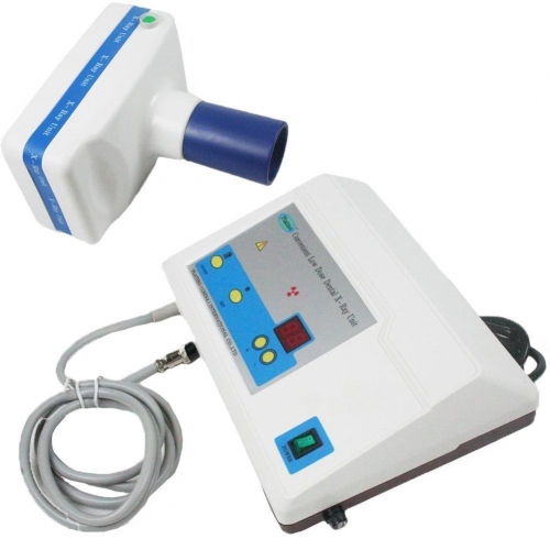
The Benefits of Digital Dental X-ray

Digital radiography is a type of X-ray imaging in which the images are transposed digitally onto computers or other devices rather than being developed onto film. Instead of using electromagnetic radiation and chemical processing to record an X-ray onto film, digital versions use digital sensors to record images onto an image capture device, which then creates a digital image file.

This file can then be used by medical staff members, and the file can be attached to a patient’s medical notes for future reference. It can be printed to paper or slide material so can be used the same as any standard X-ray, but without as much risk and usually at lower overall cost. The initial expenses with a digital system can be immense, but over time they usually pay for themselves. These sorts of imaging devices are most commonly seen in hospitals, specialist medical practices, and dental offices. Dental imaging requires a slightly different process but the technology and basic concept are the same.

Digital dental X-rays are used by your dentist to take images of your mouth, including tooth structure and your jaw bones. In order to take the digital images, your dentist – or a dental technician – will place a small sensor in your mouth, carefully positioned. This small sensor is connected to the processing computer by a very thin wire.

Your dentist or the dental tech inputs the command for the dental X-ray machine to send a X-ray through your teeth and into the sensor, effectively taking a photo of your tooth or teeth. The sensor captures the resulting image and sends it through the wire to the computer. Then your dentist will reposition the sensor and take additional digital X-rays until all of your teeth have been X-rayed.

With digital dental X-rays, your dentist or other dental professional is able to immediately see your teeth and jaw bones. This means that assessment and diagnosis is virtually instantaneous.

Digital Dental X-Rays – A Great Advance

Many dentists are now using digital dental X-rays, which have many benefits for you, as the patient. Among some of the many benefits for you, are:
1. A reduction in the amount of radiation you are exposed to during the X-ray process.
2. No need for dental film or processing chemicals in order for your dentist to be able to see the images.
3. An almost instantaneous ability to see the X-ray images, which means, if you’re in pain, you’re not waiting for the images to be processed and then viewed.
4. Your dentist is able to use color contrast in viewing and manipulating the X-ray image, in order to see all of the details in the image.

The Use of Dental X-ray Machine

In dentistry, there are two types of digital imaging systems used in intraoral radiography: computed radiography (CR) and direct radiography (DR). These are then categorized into periapical and panoramic x ray machines. Furthermore, there are two sources of image noise used in digital imaging: statistical noise and structured noise.

Dental radiography has evolved from film and chemical developers into a highly technical process that involves various types of digital x-ray machines, as well as powerful dental software programs to assist the dentist with image acquisition and diagnostic analysis of the acquired images. When making the decision to purchase x-ray equipment, the doctor needs to research the available options thoroughly, in order to make an informed choice for the “right” machine for his or her practice.

The orthodontist requires a way to obtain the size and form of craniofacial structures in the patient. For this reason, a cephalometric extension on the imaging x-ray device is necessary to acquire images that evaluate the five components of the face, the cranium and cranial base, the skeletal maxillae, the skeletal mandible, and maxillary dentition. The cephalometric attachment offers images such as frontal AP and lateral cephs.

If the practice is concentrated in endodontic and implant treatment, then a CBCT machine is the most practical method of providing the doctor with diagnostic tools such as mandibular canal location, surgical guides, and pre-surgical treatment planning with the assistance of powerful 3D dental software applications. The patient is benefited by the reduced radiation exposure provided by these machines.

Like old fashioned dental X-rays, digital dental X-rays are used by your dentist to take images of your mouth, including tooth structure and your jaw bones. In order to take the digital images, your dentist – or a dental technician – will place a small sensor in your mouth, carefully positioned. This small sensor is connected to the processing computer by a very thin wire.

Your dentist or the dental tech inputs the command for the dental X-ray machine to send a X-ray through your teeth and into the sensor, effectively taking a photo of your tooth or teeth. The sensor captures the resulting image and sends it through the wire to the computer. Then your dentist will reposition the sensor and take additional digital X-rays until all of your teeth have been X-rayed.

With digital dental X-rays, your dentist or other dental professional is able to immediately see your teeth and jaw bones. This means that assessment and diagnosis is virtually instantaneous.

The Development of Dental Radiography

Dental radiography has evolved from film and chemical developers into a highly technical process that involves various types of digital x-ray machines, as well as powerful dental software programs to assist the dentist with image acquisition and diagnostic analysis of the acquired images.

“What is the main type of treatment that I provide my patients?” If you are a general practitioner, a standard 2D panorex will provide all of the imaging requirements needed for such treatments as caries detection, diagnosis of TMJ issues, OPG images, and images of the patients entire detention in a single x-ray. Many of the newer 2D panoramic units also offer extraoral bitewing imaging capability, which allows the dentist to obtain a bitewing image without putting a sensor or periapical film inside of the patient’s mouth.

Your dental X-rays allow your dentist to see the condition of prior dental procedures, such as fillings, crowns, root canals, and bridges. And, too, your dentist will be able to look for possible bone loss as a result of periodontal gum disease and find hidden tartar build up. In addition to diagnostics, your dentist may choose to perform dental X-rays in order to check for bone density as part of preparing you for dental implants, which require an adequate density in order to support the implants.

Electromagnetic radiation has been used in the medical profession for imaging and diagnostics for many years. X-radiation technology, known more simply as X-ray technology, allows accurate images to be captured of a person, animal, or thing’s internal composition. A generator uses strong electromagnetic light paired with a detector; most things, humans included, will naturally absorb some of the light, which is what allows the detector to map out images and specific locations.

Most of the earliest X-rays depended on photographic films to capture the images and make them readable. Digital detectors skip this step; rather than using light beamed through objects onto film, it allows for digital scanning and image interpretation. In terms of radiation the two are about the same initially, though digital versions typically have a shorter exposure time and as such tend to be more efficient.

Digital radiographs are becoming a popular form of dental x-ray machines. They use an electronic pad or sensor instead of a piece of x-ray film. This kind of dental x-ray machine transmits images directly to a computer. The images can be stored, printed, and compared using software to decide whether any changes have occurred over time. A technician can view two different images or multiple images on a single screen view. Digital systems even make it possible to catch dental problems sooner than with other methods and types of dental x-ray machine.