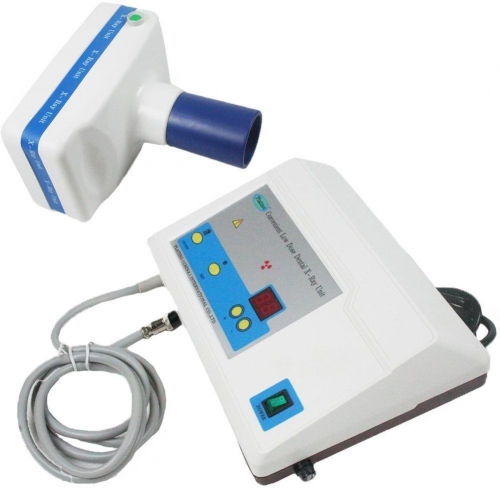
In dentistry, there are two types of digital imaging systems used in intraoral radiography: computed radiography (CR) and direct radiography (DR). These are then categorized into periapical and panoramic x ray machines. Furthermore, there are two sources of image noise used in digital imaging: statistical noise and structured noise.
Dental radiography has evolved from film and chemical developers into a highly technical process that involves various types of digital x-ray machines, as well as powerful dental software programs to assist the dentist with image acquisition and diagnostic analysis of the acquired images. When making the decision to purchase x-ray equipment, the doctor needs to research the available options thoroughly, in order to make an informed choice for the “right” machine for his or her practice.

The orthodontist requires a way to obtain the size and form of craniofacial structures in the patient. For this reason, a cephalometric extension on the imaging x-ray device is necessary to acquire images that evaluate the five components of the face, the cranium and cranial base, the skeletal maxillae, the skeletal mandible, and maxillary dentition. The cephalometric attachment offers images such as frontal AP and lateral cephs.
If the practice is concentrated in endodontic and implant treatment, then a CBCT machine is the most practical method of providing the doctor with diagnostic tools such as mandibular canal location, surgical guides, and pre-surgical treatment planning with the assistance of powerful 3D dental software applications. The patient is benefited by the reduced radiation exposure provided by these machines.
Like old fashioned dental X-rays, digital dental X-rays are used by your dentist to take images of your mouth, including tooth structure and your jaw bones. In order to take the digital images, your dentist – or a dental technician – will place a small sensor in your mouth, carefully positioned. This small sensor is connected to the processing computer by a very thin wire.
Your dentist or the dental tech inputs the command for the dental X-ray machine to send a X-ray through your teeth and into the sensor, effectively taking a photo of your tooth or teeth. The sensor captures the resulting image and sends it through the wire to the computer. Then your dentist will reposition the sensor and take additional digital X-rays until all of your teeth have been X-rayed.
With digital dental X-rays, your dentist or other dental professional is able to immediately see your teeth and jaw bones. This means that assessment and diagnosis is virtually instantaneous.