Category Archives: dental tips

How to Clear Chairside Sandblasting

An advance in adhesive dentistry has resulted in sandblasting, to increases micro-retention, being performed as a routine procedure. Instead of wearing a path from the patient’s portable folding chair to the office lab to clean excess cement from a patient’s temporary or loosened permanent crown ,or for sandblasting the fitting surface of a crown, bridge inlay or veneer, the procedure is a half- turn away, thanks to the new breed of sandblasters and hookup options.

The uninterrupted patient/doctor exchange is especially beneficial with anxious adult patients – no need to cut the reassuring golf story short for a trip down the hall, leaving the patient alone. Standard hookup kits allow, with a simple male disconnect, access to the dental unit’s air source through the female port.

Many dentists have sandblasters with quick disconnects in every operatory, and these space- efficient wonders tuck easily into a drawer. The adaptors for standard 4-hole dental handpiece ports,or even for your favorite Kavo®, Sirona® or W&H® High speed handpiece port, for a blasting procedure – just pop on the adaptor and activate the sandblaster with your regular foot control, And how about the air quality?

The old dust collection methods are fast disappearing as dentists perform more and more chairside sandblasting. You’ve heard of them as the homemade variety, consisting of a discarded packaging box of gallon plastic milk carton with three handcuts holes. Learning over the nearest trash can, much to the dismay of office staff, Best of all, throwing open a window and blasting away (weather permitting). Times have changed and the new waves of dust collectors not only keep the air clean, but they look great, too – they’re high-tech looking, lightweight and simple to operate and empty.

No fancy installation, either- they simply plug into the nearest outlet. Best of all, the new breed of dust collectors are scaled to fit comfortably on an operatory countertop without without getting in the way, and without compromising user comfort or efficiency.
The Uses of Dental Air Polisher

Use of dental air polishers decreased as the clinician’s age and experience increased. Curricula in many dental hygiene schools do not include clinical instruction in the use of this polishing device due to inadequate numbers of units and difficulty in moving units between clinic stations. Inadequate or insufficient knowledge and experience, therefore, appears to be a major factor in the underutilization of the air polisher.

In an attempt to provide a suitable knowledge base for practicing dental hygienists, the primary purpose of this article is to provide a comprehensive summary and critique of the research on all aspects of air polishing. In addition, a suggested technique, common concerns, and possible solutions will be discussed.

Discussions are based on a review of the relevant literature on air polishing. Tables organize the data into categories to facilitate access of needed information. Because of the various research designs employed and the number of variables that must be controlled, comparative analyses of the studies are difficult. However, where possible, analyses of the validity and reliability of the studies are provided. It should be remembered that while laboratory (in vitro) investigations are useful, the most definitive conclusions must be obtained through clinical (in vivo) studies. Case reports or opinion articles have limited applications.

Because polishing with a rubber cup and prophylaxis paste has been shown to remove the fluoride rich layer of enamel and cause marked loss of cementum and dentin over time, this method of stain removal has been challenged Rubber cup polishing with prophylaxis pastes, therefore, may not be a suitable method for moderate-to-heavy stain removal on enamel, cementum, or dentin. One study, however, contradicted these findings and suggested that rubber cup polishing with chalk is equally effective in decreasing root-surface roughness caused by sonic scaling. Chalk is not a common polishing agent and no comparison of the abrasiveness was made between it and the sodium bicarbonate used in air polishing powder. This study, therefore, may not simulate actual clinical situations.

Numerous investigations have examined the effects of the air polisher on a variety of restorative materials. Some results have been positive, while others have recommended caution near restorations. Although some studies are contradictory, most suggest caution or complete avoidance when air polishing on or near composite restorations. On composites, surface roughness or pitting was the most common result seen. One study concluded that, although marginal microleakage was greater for composites than for amalgams, this loss was not statistically or clinically significant. More research is recommended since previous studies do not support this conclusion.

The Tips for Choosing Dental X-ray Machine

Finding the right technology for a practice requires research, investment and care. As more dental professionals implement digital intraoral radiography( intraoral camera ) into their practices, dental assistants should be sure to not only become educated on proper use, but also proper care.

When contemplating the change to digital dental in your practice, the choices can be confusing for the dentist. Dental radiography has evolved from film and chemical developers into a highly technical process that involves various types of digital x-ray machines, as well as powerful dental software programs to assist the dentist with image acquisition and diagnostic analysis of the acquired images.

When making the decision to purchase x-ray equipment, the doctor needs to research the available options thoroughly, in order to make an informed choice for the “right” machine for his or her practice.

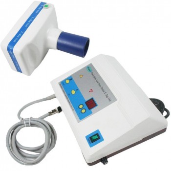
While many patients see their dentist in-office, others require the dentist and equipment to go to them. Those who are incarcerated, home-bound, in nursing homes, working in underdeveloped locations or stationed on military bases are just some of the patients who may benefit from having access to a portable x-ray machine. Teeth problems could not only be painful but could also cause many health problems. Waiting to access an in-office machine may not be an option depending on the condition.

The orthodontist requires a way to obtain the size and form of craniofacial structures in the patient. For this reason, a cephalometric extension on the imaging x-ray device is necessary to acquire images that evaluate the five components of the face, the cranium and cranial base, the skeletal maxillae, the skeletal mandible, and maxillary dentition. The cephalometric attachment offers images such as frontal AP and lateral cephs.

The portable dental x-ray is not only useful to patients, but also to dentists who want to be able to help patients who don’t have immediate access to a dental office. Without the device, there is no doubt that quite a few individuals would go without knowing the cause of their tooth pain. Though it doesn’t mean they’ll seek immediate dental care, it at least increases the chances depending on the results of the x-rays.

How Can You Save Money on Dental Equipment

Operating a successful dental practice is not exactly cheap, but there is a litany of ways that you can save money along the way. Find a trusted dental equipment resource online and stop overspending for important equipment.

Finding the best equipment possible should be your top priority, you also need to consider the financial impact of these investments. Here are three important pieces of dental equipment that you should never overspend on:

One of the biggest expenses you will incur as a dentist is the money you spend on replacement parts. From compressor parts to dental bulbs, you’ll find yourself in need of replacement equipment on a weekly basis. About 74% of adults believe an unattractive smile can hurt their success, and showing your patients “unattractive” and worn-down equipment will certainly hurt your success moving forward. Always try to keep these replacement parts as new as possible without exceeding your budget.

Dental autoclaves are truly the backbone of any dental office, and you cannot work safely without them. All of your handheld tools need to be sterilized before they can be used, and you can end up spending way too much on these devices if you invest in a lower-tier product that constantly needs to be replaced. Searching online will allow you to find dental autoclaves that suit your specific needs, including automatic autoclaves, manual devices, and chemical autoclaves.

Dental cone beams are a fairly new technology that has revolutionized the way that dentists can treat their patients. When regular facial x rays are not sufficient, you need an advanced tool that will allow you to diagnose urgent dental issues. There are two types of digital imaging systems used in intraoral radiography(intraoral camera) — computed radiography (CR) and direct radiography (DR). Purchasing dental cone beams online enables you to pick the machine that is right for your practice and avoid the dreaded “buyer’s remorse.”

Opening a dental practice is not an inexpensive task, and you’ll find yourself spending more and more money over time to sustain your success. Therefore, you should always be on the lookout for high-quality equipment at an affordable price.

What Should You Know about Endodontic Treatment

Endodontics is a specialized type of dentistry that deals with abnormal tooth pulp; the causes/origins, diagnosis, prevention and treatment of diseases and injuries to the pulp; and other conditions affecting the tissue surrounding the pulp and canal (channel inside the tooth root).

Depending on your situation, your dentist or endodontist may perform any one or combination of procedures to treat your condition. The cost for endodontic therapy typically averages $600 to $900. The cost for a therapeutic pulpotomy (excluding final restoration) is approximately $150, and the cost for apicoectomy/periradicular surgery ranges from $550 to $700.

Endodontic procedures are used in the diagnosis and treatment of oral pain involving the pulp and periradicular area (just outside or around the root of the tooth origin). Pulp therapy, such as pulpotomy, is a common endodontic procedure in which dental pulp tester is removed from the pulp chamber. The nonsurgical treatment of root canals, especially in difficult cases such as teeth with blocked, narrow or unusually positioned canals, also is a major part of endodontic therapy.

Endodontic treatment may also be required for surgical removal of diseased or abnormal (pathologic) tissues, repair procedures associated with the surgical removal of pathologic tissues, repair of cracked teeth or the replacement (replantation) of teeth knocked out (avulsed) by injury.

Surgical removal of tooth structure, such as an apicoectomy, or root-end resection (the removal of the root tip and the surrounding infected tissue of an abscessed tooth), hemisection (the process of cutting a tooth with two roots in half) and bicuspidization (procedure to change tricuspid valve into a functioning bicuspid valve).

Endodontic implants, which extend through the root canal into the periapical bone structure (tip or apex of the root of a tooth), whereas other types of tooth implants are anchored directly in the gums or jawbones.

If a root canal procedure is not performed, an abscess (infected pus pocket) can form at the tip of the tooth root that can be painful. Even if there is no pain, the bone holding the tooth in the jaw can be damaged.
When is Endodontic motor Treatment Appropriate?

Endodontic treatment is necessary when the dental pulp becomes inflamed or infected as a result of deep tooth decay, repeated dental procedures on the tooth or a crack, chip or other injury to the tooth. Most teeth can be saved by endodontic treatment.

Q. Why can’t smokers undergo certain cosmetic dentistry procedures?

A. Smoking is a leading cause of tooth loss. Smokers also have an increased risk for periodontal disease (gum disease), leukoplakia, loss of bone structure, inflammation of the salivary gland, and development of lung, throat, or oral cancer. Smokers therefore face an additional set of considerations when exploring cosmetic dentistry treatment.

One restriction for smokers involves dental implants. Smoking increases the risk that a dental implant will fail to integrate with bone tissue (osseointegration). Smoking also affects gum health, and unhealthy gums can lead to dental implant failure. If you smoke, you need to quit before having an implant.

Another restriction involves teeth whitening. If you whiten your teeth but continue to smoke, the whitening effect will not last as long, because smoking stains the teeth.

Q. May I choose to be sedated during cosmetic dentistry procedures?

A. Yes. The level of sedation necessary for cosmetic dentistry procedures depends on several factors, including:

Your health and medical history.
The procedure being performed.
Your dentist’s familiarity with sedation modalities.

Most people do not require a high level of sedation during cosmetic dental procedures.

Q. Are dental implants suitable for children?

A. Dentists indicate that implants cannot be considered until bone growth has completed. This typically occurs during adolescence, but may vary from one child to the next. Implants placed prior to bone growth completion may shift out of place and cause severe problems. If a child loses a tooth before bone growth has completed, an orthodontic device called a “space maintainer” may be used to prevent an abnormal growth of surrounding teeth. Once bone growth has completed, the space maintainer can be removed and the missing tooth restored using an implant or bridge.

Q. Are veneers my only option if I want a smile makeover?

A. No. Your cosmetic dentist will determine the cosmetic dentistry procedures that would be the best for you. The dentist’s suggestions will be based on your desires and a thorough examination to determine the condition of your teeth and what problems, if any, may require dental treatment. Therefore, your smile makeover could involve teeth whitening, crowns, composite bonding, gingival sculpting, inlays and onlays or any other combination of cosmetic dental procedures that your dentist determines is right for your cosmetic treatment plan.

Q. Does led teeth whitening work for everyone?

A. No. In cases involving severe discoloration, teeth whitening may not result in a bright, white smile. Individuals with severe tooth discoloration should consider other options, such as porcelain veneers or composite bonding.

For more information, please visit: https://www.alandental.com/category-55-b0-Portable-X-ray-Machine.html

What Should You Pay Attention on the Procedure of Cosmetic Dentistry

Although dentures are not considered a cosmetic dentistry procedure, they provide a cosmetic benefit for people who have lost all or some of their teeth. People who are missing only certain teeth, or rows of teeth, may be better served by a dental bridge or dental implants. It is best to have your situation evaluated by a dental professional in order to establish candidacy for any of these procedures.

The level of sedation necessary for cosmetic dentistry procedures depends on several factors, including:

Your health and medical history.
The procedure being performed.
Your dentist’s familiarity with sedation modalities.

Most people do not require a high level of sedation during cosmetic dental procedures.

Smoking is a leading cause of tooth loss. Smokers also have an increased risk for periodontal disease (gum disease), leukoplakia, loss of bone structure, inflammation of the salivary gland, and development of lung, throat, or oral cancer. Smokers therefore face an additional set of considerations when exploring cosmetic dentistry treatment.

One restriction for smokers involves dental implants. Smoking increases the risk that a dental implant will fail to integrate with bone tissue (osseointegration). Smoking also affects gum health, and unhealthy gums can lead to dental implant failure. If you smoke, you need to quit before having an implant.

Another restriction involves led teeth whitening. If you whiten your teeth but continue to smoke, the whitening effect will not last as long, because smoking stains the teeth.

some children and adolescents may have severely discolored teeth as a result of taking certain medications. Or, the front teeth may have been fractured due to an accident or sports injury and require composite bonding or veneers. In other instances, there still may be slight gaps or spaces between the teeth even after orthodontics, so perhaps composite bonding would enhance the look of the smile.

If your child or teenager is concerned about the appearance of his or her smile, make an appointment to see your dentist. He or she will suggest the most appropriate course of action based on your child’s age, oral condition and desires, as well as possibly refer you to a cosmetic dentist for consultation.

How Can You Know Dental Handpiece not Work Properly

The dental handpiece is the most used piece of equipment in any dental practice today. This piece of equipment is used in almost every dental procedure, so recognizing the signs that your handpiece isn’t working properly is critical to the productivity of your practice and the comfort of your patients.

The air driven handpiece is still the mainstay within the industry, but electric handpieces are being used more and more as technology advances. The newest technology takes components from both air driven and electric handpieces.

Understanding how to properly clean and maintain these handpieces, as well as their components, will help the clinician achieve optimal results. You should clean your
dental handpiece with a soft bristle brush & warm water before autoclaving to remove any organic material that might corrode your handpiece. It is not advised to use detergents or soaps, as they can destroy the optics, get into the bearings & weaken the lubrication, which can reduce the life of your dental handpiece.

Reduction in Speed: Over time, especially if the handpiece is used a lot, the rotation speed decreases. This is caused by a number of things, but the most common is a faulty turbine. This sign is important, because the loss of speed in your handpiece will prevent you from performing timely and successful treatments.

Vibrations: If your handpiece starts to vibrate, then this can be an indication the bearings are wearing out or loose. Additionally, this problem can be caused by lack of lubrication. If this is the case, you will need to have the handpiece repaired by a handpiece repair specialist.

Disturbed rhythm: If your handpiece makes clunking noises and rotates irregularly, this could be a motor or turbine problem. This is often caused by old age or a lack of regular servicing, and you will need to get your brushless micro motor or turbine replaced. This will ensure consistently smooth revolutions that don’t pose such a risk to the teeth and gums of your patients.

Makes Noise: If your handpiece starts makings a loud chunking noise, this is an indication that the handpiece was probably dropped and the head was dented. Once this happens, the impeller within the turbine is compromised due to the drop. When the impeller within the turbine isn’t rotating smoothly the turbine will fail causing the bur to slip.

If you notice any of these signs with your dental handpieces, it is important to quickly find a reputable handpiece repair company to help protect your investment, minimize your handpiece downtime and keep your handpieces running at optimal performance.

The Most Critical Step in Root Canal Therapy

In the past, access cavities were standardized mainly dependent on the tooth type. However, with the advent of modern endodontic motor techniques using a dental operation microscope & loupes providing magnification & better illumination, the ‘ideal’ access cavity preparation has evolved from being based on individual tooth type to the preparation based on the shape of the pulp chamber morphology of the tooth being treated.

Access is the first & arguably the most important phase of non-surgical root canal treatment. However, it can be the most challenging and frustrating aspect of endodontic treatment. Therefore, for successful treatment, good access cavity design and preparation is imperative for quality endodontic treatment, prevention of iatrogenic problems & prevention of endodontic failure.

As novices to root canal therapy ourselves, one of the worst nightmares during access cavity preparation is perforation. Furthermore, there are several aspects of access cavity preparation that could go wrong without the correct knowledge & guidance that plagued us during our graduation days. Now, however, being armed with the correct knowledge & guidance, we can recognise the cause & prevent these procedural errors during treatment by following a set of few simple laws & guidelines that govern & dictate the access cavity preparation.

Access into the pulp chamber and the root canal system is the most critical step in root canal therapy. Unlike other aspects of Dentistry, root canal therapy is carried out with limited visual guidance. This, coupled with complex anatomy of the root canal system & various pathological conditions, makes the preparation of an ideal access a crucial step to gain success in treatment. Therefore, the objective of a well designed access cavity is to create a smooth, straight line path to the root canal system, while retaining as much tooth structure as possible.

Therefore, the purpose of this article is to highlight the common iatrogenic errors by General Dental Practitioners due to the lack of knowledge & awareness of certain internal anatomic features for a given tooth.

For more information, please visit: https://www.alandental.com/category-82-b0-Dental-Pulp-Tester.html

The Technique of Imaging in Endodontics

CT in addition to 3D images offer several advantages over conventional radiography: it eliminates anatomical noise and high contrast resolution, allows differentiation of tissues with less than 1% physical density difference to be distinguished in comparison to conventional radiography that requires 10%. When examining jaws, axial scans are usually acquired to avoid artifacts caused by posts, crowns, and metallic fillings .

CT can even supply additional information about the morphology of the root-canal system provided that it does not contain metallic root-canal posts . However, the geometric resolution of CT is insufficient to reveal the exact shape of the root-canals , and a very high radiation dose is required to achieve a high enough resolution to assess root-canal anatomy in detail .CT may also be useful for the diagnosis of poorly localized odontogenic pain. In some circumstances in which periapical radiographs reveal nothing untoward, CT may confirm the presence of a periapical lesion .

The assessment of the ‘third dimension’ with CT imaging allows the determination of the number of roots and root canals,as well as where root canals join or divide. This knowledge is useful when diagnosing and managing failed endodontic motor treatment. CT can also be used to localize foreign bodies in the jaws such as gutta-percha and root-canal sealer. CT in endodontics has however some disadvantages such as the high radiation dose and the high costs of the scans . Other disadvantages are scatter due to metallic objects, relatively low resolution in comparison to conventional radiographs.

Micro-computed tomography (micro-CT), another alternative CT technique, has been considered in endodontic imaging .The use of micro-CT remains a research tool limited to in vitro measurementsof small samples; due to the high radiation dose required, and cannot be employed for human imaging in vivo .

Ultrasound imaging is based on the reflection of sound waves (echoes), with a frequency outside the rangeof human hearing (1-20 kHz), at the interface of tissues which have different acoustic properties . The echoesare detected by a transducer which converts them into an electrical signal, and a real-time black, white and shades of grey echo picture is produced on a computer screen .

The technique is easy to perform and may show the presence, exact size, shape, content and vascular supply of endodontic lesions in the bone . Ultrasound has been found to be a reliable diagnostic technique in the differential diagnosis of periapical lesions (granulomas versus cysts) with the aid of the echo picture (hyperechoic and hypoechoic) and through the use of the colour laser Doppler effect to provide evidence of vascularity within the lesion .

See more: https://www.alandental.com/category-82-b0-Dental-Pulp-Tester.html