All posts by admin_alan

The Use of Digital Tech in Dentistry

When contemplating the change to digital dental in your practice, the choices can be confusing for the dentist. Dental radiography has evolved from film and chemical developers into a highly technical process that involves various types of dental x-ray machines, as well as powerful dental software programs to assist the dentist with image acquisition and diagnostic analysis of the acquired images. When making the decision to purchase x-ray equipment, the doctor needs to research the available options thoroughly, in order to make an informed choice for the “right” machine for his or her practice.

Digital radiography is a type of X-ray imaging in which the images are transposed digitally onto computers or other devices rather than being developed onto film. Instead of using electromagnetic radiation and chemical processing to record an X-ray onto film, digital versions use digital sensors to record images onto an image capture device, which then creates a digital image file. This file can then be used by medical staff members, and the file can be attached to a patient’s medical notes for future reference.

Dental X-rays are one of the most important part of your regular dental treatment. You use the specialized imaging technology to look for hidden tooth decay – also called cavities – and can show dental issues such as abscessed teeth, dental tumors, and cysts.

While many patients see their dentist in-office, others require the dentist and equipment to go to them. Those who are incarcerated, home-bound, in nursing homes, working in underdeveloped locations or stationed on military bases are just some of the patients who may benefit from having access to a portable dental x-ray. Teeth problems could not only be painful but could also cause many health problems. Waiting to access an in-office machine may not be an option depending on the condition.

If the practice is concentrated in endodontic and implant treatment, then a CBCT machine is the most practical method of providing the doctor with diagnostic tools such as mandibular canal location, surgical guides, and pre-surgical treatment planning with the assistance of powerful 3D dental software applications. The patient is benefited by the reduced radiation exposure provided by these machines.

What Should You Know about the Dental Air Compressor

Safety should always be your first priority when working with compressed air, no matter the setting. It may seem like a simple tool and you might be tempted to blast a coworker with a burst of air, or use the compressed air to blow dirt or dust away from your skin, but this is dangerous behavior.

Compressed air is under such pressure that even brief contact with protected skin can cause abrasions, cuts and other injuries. If the skin is broken, an air compressor can force air under the skin, causing organ damage or even potentially causing an embolism — an air bubble in the blood that can cause a cardiac event if it reaches the heart. In rare cases, impacts from compressed air have been known to cause traumatic organ damage.

An air compressor could have perfectly functioning filtration and be free of condensate or traveling oil, yet still lag in its performance if air leaks are present at any point between the machine itself and the tools at the end of the pipes.

When air leaks form along a compressed air system, tool performance weakens and operations become less efficient. If an operator is unaware of the problem source, the first solution that often comes to mind is to simply crank up the pressure on the system in order to compensate for the lagging power. This, in turn, leads to increased wear and tear on the internal mechanisms of an air compressor.

Simply put, there’s nothing to be gained from leaks in a compressed air system. The problems associated with leaks are easier to spot and remedy, or prevent altogether, with routine inspections along all the parts that transport air from the compressor to the end tools.

Compressors need lubrication to function, but the oil can get into the airstream, where it could threaten patient health and jeopardize procedures. Some units are oil-free, while others have special sealant systems to prevent leaks. The dental air compressor may also be designed to operate quietly, which can reduce stress for patients who may be worried by the sound of a large engine running near the procedure room.

Buying a name-brand compressor can help reduce that risk even further by giving you access to approved service providers who are trained to maintain your specific piece of equipment. While using a different service provider for your dental equipment maintenance is one option, you aren’t guaranteed to be paired with a technician who knows all the ins and outs of your compressor’s idiosyncrasies.

How to Use Air Polishers Effectively

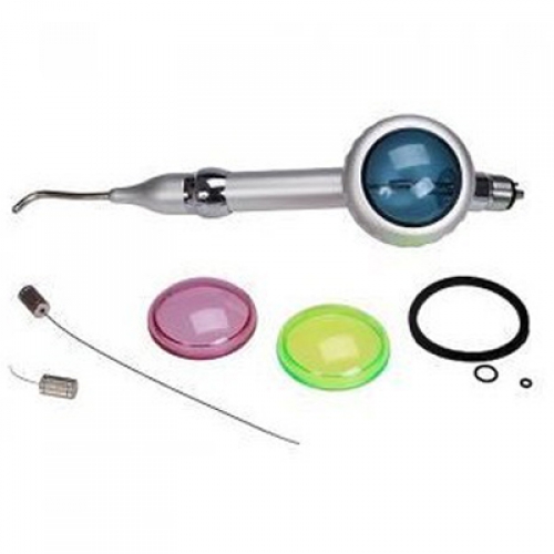
Use of the dental air polisher for stain removal involves three steps: patient selection and preparation, clinician preparation, and the actual clinical technique. Air polishing should follow a careful review of the patient’s medical and dental history, and a thorough examination of the oral hard and soft tissues. Indications and contraindications, effects on hard tissues, restorations, safety, and alternative uses should be reviewed prior to treatment planning the use of the air polisher.

Preparation of the patient should include an explanation of the procedure, removal of contact lenses, an anti-microbial rinse, application of a lubricant to the lips, placement of safety glasses or a drape over the nose and eyes, and placement of a plastic or disposable drape over the patient’s clothing. Operators should use universal precautions, including protective apparel, a face shield or safety glasses with side shields, gloves, and a well-fitting mask with high-filtration capabilities.

During periodontal surgery, air polishers can prepare root surfaces detoxify them effectively and efficiently, and leave a uniformly smooth root surface that is clean and free of diseased tissues. Dentinal tubules are then occluded, which may result in decreased sensitivity. Superior growth and vitality of human gingival fibroblasts was evident when ultrasonic scaling was followed by air polishing, compared to ultrasonic scaling alone. Air polishing produced root surfaces that were comparable to manually rootplaned surfaces, and provided better access to furcations. Tissue healing following air polishing was comparable to that achieved by hand instrumentation in root preparation during periodontal flap surgery.

Research findings also support the use of air polishing with orthodontic patients. It is the most efficient and effective method for plaque and stain removal around orthodontic brackets, bands, and arch wires. It is not contraindicated on orthodontic bracket composite resin adhesive systems.

In restorative dentistry, air polishers have provided stronger composite repairs than traditional etching gels. They also are superior to rubbercup polishing in preparing occlusal surfaces for etching prior to sealant placement because the rubber cup forces debris into the fissures. Air polishing of occlusal surfaces also allows for deeper penetration of the sealant resin into the enamel surface than rubber cup and pumice cleaning of the fissures. Air polishers also have enhanced sealant bond strength compared to traditional polishing with a low-speed handpiece, bristle brush, pumice, and water.

Oral health care professionals have a responsibility to patients to engage in life-long learning in order to provide the most contemporary clinical care. Air polishing has been studied extensively and, when used appropriately, provides a safe, efficient and contemporary approach to achieving a variety of treatment goals.

The Information of Dental Root canals

It’s been said that root canals hurt, that they cause disease, and that you only need to get one when a tooth is painful. One myth even alleged that root canals cause cancer and other diseases. With stories like that swirling around the world of dental care, it’s no wonder people dread root canals. But there’s good news! All those myths have been busted. So if your dentist sits you down to talk about when to get a root canal, don’t run for the door.

Every tooth consists of three different layers. The outermost and hardest layer is enamel, and the second layer is dentin. The third is pulp, which is the cavernous space where the live tissue and nerve of each tooth is located.

If for any reason the pulp space( pulp tester ) is exposed to the outside, the tissue becomes contaminated and eventually infected. The exposure of pulp happens in many circumstances, such as when you have a large cavity or a fractured tooth. Your dentist can explain the exact reason for damage to this tissue. In these cases, the treatment is usually root canal treatment.

Your teeth are strong, but poor dental care can cause the protective enamel that encases your teeth to break down. When this happens, your teeth become sensitive and more vulnerable to fractures and cavities. This is when your tooth’s root can become infected. Each tooth has soft tissue that runs through the tooth’s canals. The tissue is made up of nerves and blood vessels, which provide vital nourishment to the tooth. When a cavity or fracture is left untreated, that pulpy, soft tissue becomes infected and causes pain – that’s when to get a root canal.

Root canals are a common procedure used to save your tooth once infection sets in. If untreated, the infection can spread and cause damage to surrounding bone. Once this happens, there’s a greater risk of losing your tooth.

To remove the infection and stop it from spreading, a small opening is created at the top of the tooth. From this opening, the tooth’s nerve is removed from the canal, and the inside of the tooth is cleaned before the canals are filled with a rubber-like material. Filling in the now-empty canals helps to seal them from future infection. Once complete, a temporary filling is placed onto the tooth until a permanent filling or crown is ready.

Swelling around your gums and lips will last for a few hours after the root canal, and lingering swelling can last for a few days. Don’t be alarmed if your tooth is still sensitive. This is normal following a root canal, but talk with your dentist about what to expect during recovery. Avoid chewing hard foods until a permanent crown is put on, and talk with your dentist about long-term care for the afflicted tooth. Root canals have a high success rate, but like all of your teeth, they can be damaged if they are not taken care of properly. Good oral hygiene and regular visits to the dentist are key to maintaining the condition of your teeth whether you’ve had a root canal or not.

The Benefits of Dental Air Polishing

Like many things in dentistry, air polishing has changed since the late 1970s. There are several new dental air polisher designs and new abrasive powders, and we now have the ability to use air polishers subgingivally. Sodium bicarbonate was the original abrasive powder used, but newer abrasive powders include glycine, calcium carbonate, calcium sodium phosphosilicate, aluminum trihydroxide, and erythritol. These additional powders give us more choices for our patients’ individual needs.

Use of air polishers decreased as the clinician’s age and experience increased. Curricula in many dental hygiene schools do not include clinical instruction in the use of this polishing device due to inadequate numbers of units and difficulty in moving units between clinic stations. Inadequate or insufficient knowledge and experience, therefore, appears to be a major factor in the underutilization of the air polisher. In an attempt to provide a suitable knowledge base for practicing dental hygienists, the primary purpose of this article is to provide a comprehensive summary and critique of the research on all aspects of air polishing. In addition, a suggested technique, common concerns, and possible solutions will be discussed.

Discussions are based on a review of the relevant literature on air polishing. Tables organize the data into categories to facilitate access of needed information. Because of the various research designs employed and the number of variables that must be controlled, comparative analyses of the studies are difficult. However, where possible, analyses of the validity and reliability of the studies are provided. It should be remembered that while laboratory (in vitro) investigations are useful, the most definitive conclusions must be obtained through clinical (in vivo) studies. Case reports or opinion articles have limited applications. Therefore, interpretation and application of research results must be done with caution.

The benefits of air polishing for the dental professional include less operator fatigue, less time involved than the traditional polishing technique, and improved access to difficult-to-reach areas. Benefits to the patient include less time in the chair, less “scraping,” excellent stain removal, reduced dentin hypersensitivity, and improved periodontal status.

Air polishing has been compared to scaling( dental scaler ) and rubber-cup polishing for efficiency and effectiveness of stain and plaque removal. The use of the air polisher as an efficient and effective means of removing extrinsic stain and plaque from tooth surfaces. Air polishing requires less time than traditional polishing methods and removes stain three times as fast as scaling with comers. In addition, less fatigue to the operator has been mentioned as an important benefit of air polishing.

Why You Need Dental Autoclaves

Effective and efficient infection control in the dental office is essential for the safety of patients and to ensure that productivity does not suffer. Infection control programs all include the cleaning and sterilization of reusable dental instruments and devices.

One of the dental equipment, dental autoclaves, plays a crucial role in keeping your patients healthy. Since autoclaves are involved in the sterilization of dental instruments, you simply cannot afford to invest in anything but the best. However, you should know that there are several varieties and price points of autoclaves available to suit your specific needs.

About 74% of people believe that an unattractive smile can hurt their career success, and a non-sterilized instrument will set them back even further. You cannot put your career on the line by failing to obtain the right kind of dental autoclave. The first thing you need to consider is that manual vs. automatic. Manual autoclaves are generally less expensive to buy and repair, but they take much longer to use. Automatic autoclaves cost a bit more, but they can save you tons of time and detect any malfunctions.

Maintain sterilized instruments in the pouches or wrapping in which they were sterilized. If the packaging becomes torn or wet, the items must be repackaged and heat sterilized. Avoid mingling non-sterile packages with sterile ones. There should be a visible indicator, such as chemical indicators or color-change autoclave tape on the outside of each package to allow staff to easily discern sterilized instrument packages from those that have not yet been heat-processed.

Sterilization is a necessary part of the maintenance of your dental handpiece. This process also puts your handpiece under the most stress with wear and tear. Maximum temperatures in your autoclave shouldn’t reach more than 140 degrees Fahrenheit. This can help to preserve your handpiece just a bit longer in between the need for service.

The same sets of instrument processing, personal protection equipment, and engineering and work practice control precautions can be expected to protect against all bloodborne disease agents. As such, instruments used on a known hepatitis patient need not be segregated from other contaminated instruments, can be cleaned in an ultrasonic cleaner, and do not require special post-cycle maintenance or cleaning of ultrasonic equipment.

You need to find that perfect balance of quality and cost to reach your full potential. Find a trusted dental equipment retailer online and find the dental autoclaves that work best for your current needs.

The Importance of Dental X-ray Machine

Your dentist uses the specialized imaging technology to look for hidden tooth decay – also called cavities – and can show dental issues such as abscessed teeth, dental tumors, and cysts. And your dental X-rays allow your dentist to see the condition of prior dental procedures, such as fillings, crowns, root canals, and bridges. And, too, your dentist will be able to look for possible bone loss as a result of periodontal gum disease and find hidden tartar build up.

In addition to diagnostics, they may choose to perform dental X-rays in order to check for bone density as part of preparing you for dental implants, which require an adequate density in order to support the implants.

The orthodontist requires a way to obtain the size and form of craniofacial structures in the patient. For this reason, a cephalometric extension on the dental x-ray machine is necessary to acquire images that evaluate the five components of the face, the cranium and cranial base, the skeletal maxillae, the skeletal mandible, and maxillary dentition. The cephalometric attachment offers images such as frontal AP and lateral cephs.

If the practice is concentrated in endodontic and implant treatment, then a CBCT machine is the most practical method of providing the doctor with diagnostic tools such as mandibular canal location, surgical guides, and pre-surgical treatment planning with the assistance of powerful 3D dental software applications. The patient is benefited by the reduced radiation exposure provided by these machines.
While many patients see their dentist in-office, others require the dentist and dental equipment to go to them. Those who are incarcerated, home-bound, in nursing homes, working in underdeveloped locations or stationed on military bases are just some of the patients who may benefit from having access to a portable dental x-ray. Teeth problems could not only be painful but could also cause many health problems. Waiting to access an in-office machine may not be an option depending on the condition.

When contemplating the change to digital dental in your practice, the choices can be confusing for the dentist. Dental radiography has evolved from film and chemical developers into a highly technical process that involves various types of digital x-ray machines, as well as powerful dental software programs to assist the dentist with image acquisition and diagnostic analysis of the acquired images. When making the decision to purchase x-ray equipment, the doctor needs to research the available options thoroughly, in order to make an informed choice for the “right” machine for his or her practice.

The Advantages of Dental Curing Light

In the dental field, a curing light can use ultraviolet or visible light, depending on what it is designed for. Both dentist and patient need to wear eye protection to limit damage to the retina for even the 20 seconds to a minute that the light is in use during rapid curing, and the light needs to be well maintained so that it will work properly and effectively. It’s also important to use the right curing light for the right resin product; many lights are designed to handle a range of resins safety.

Both light intensity – or irradiance – and the dental application should factor into a dentist’s decision regarding his or her choice of curing light. For instance, irradiance is measured by calculating power output, or milliwatts (mW), of a curing light across the surface area of the curing light guide. A curing light must deliver a minimum irradiance of 400mW/cm2 for a time interval to adequately polymerize a 1.5-2mm thick resin composite.

Clinicians also should consider the clinical application at hand. It has been documented that irradiance of curing lights attenuate/decrease significantly when it passes through restorative materials, such as ceramic restorations or resin composites. The percentage of decrease in irradiance depends on filler type, filler loading, shades, refractive index, opacity, translucency and thickness of restorative materials. Curing lights with high irradiance compensate for the decrease in the loss of total energy and allow dentists to cure resin composites completely. In general, an irradiance of 1000mW/cm2 or higher is considered ideal to cure resin-based materials through indirect restorations.

Using a curing light accomplishes two things. In the first place, it makes sure that the resin cures properly and adheres evenly. When applying fillings, this is critical to keep the filling in place in the mouth. For sealants, the curing light limits the risk of cracks and other problems with the sealant. With adhesives for implants and braces, the rapid, even cure is also designed to limit problems in the future.

The dental curing light also increases patient comfort by rapidly curing resins so that the patient is not forced to sit in discomfort while the resin sets. Since the mouth usually needs to be held open wide and may be dry for the procedure, patients usually want the procedure to end as quickly as possible so that they can close their mouths and remoisturize the dried oral membranes. Using a curing light gets patients in and out of the chair quickly so that the experience of irritation and pain is limited.

The Problems of Dental Air Compressor

One major problem that emerges in compressed air systems is pressure drop, which is marked by a loss of pressure between the compressor and the end point. While a certain amount of pressure drop is inevitable, it should never amount to more than a 10 percent loss of pressure during a given application. Otherwise, higher pressure demands end up being placed on the compressor, which results in more rapid wear and tear across the entirety of an air system.

For each pound of pressure either increased or decreased, a power plus or minus 0.5 percent is required. For example, a five percent power savings can be gained with a decrease of 10 psig. On a 100 hp compressor, this would translate to $1,740 in annual energy savings.

Pressure drop is usually caused by trouble with the pipes and weaknesses at the filters and dryers. Unfortunately, system operators will often compensate for the pressure loss by boosting the system pressure, which results in costlier operations. The correct way to handle this issue is to check for problems at the filters and dryers and replace certain items as necessary.

It’s crucial for air system pipes to remain clean and free of dirt, rust or other contaminants. After all, compressed air reaches its end point through these pipes. When contaminants are present, air pressure weakens, and the problem gradually accelerates when left un–rectified. The pipes should always remove air from the top of the air line, otherwise contaminants will travel to the pneumatic tools.

Airstream contamination increases with velocity, which in turn increases with restrictions in pipe size. Basically, the velocity of the pipes that run to the end point should be 50 seconds or less, while the interconnecting pipes and main headers should have velocity in the range of 20 to 30 feet per second. System velocity can be calculated by dividing the flow in cfm by the pipe’s compression ratio, divided by the pipe area, divided by 60.

Another factor that can impact condensation is the ambient temperature that surrounds a facility. Simply put, condensation levels multiply as temperatures rise from average to humid. For example, a 200 hp compressor will produce roughly 50 gallons of condensate over the course of a 60–degree day. However, that same machine will generate more than five times the amount of condensate if the temperature is 30 degrees higher. A 200 hp compressor will produce roughly 50 gallons of condensate over the course of a 60-degree day.

Further inefficiency can stem from problems with the system drains, which are designed to deal with condensation. Drains are placed at strategic points along a dental air compressor system, such as the tank, dryer and aftercooler. The trouble emerges when the drain fails to do its job properly, and sludge accumulates along the drainage points from a mix of water, oil and dirt.

Some of the most common mistakes in compressed air maintenance are easily avoidable if everyone on an air system staff understands how to properly care for the equipment at hand. From the compressor itself to the various parts that make up an air system, routine inspections along key areas must be made for signs of wear, condensation and dirt deposits.

The Use of Dental Intraoral Camera on Oral Pathology

The dental equipment is constantly searching for ways to improve how we can diagnose and treat conditions in the oral environment. The ability to diagnose various types of oral pathology in their earliest forms is a huge benefit to both patients and dentists.

The dental explorer is said to have an accuracy of about 25%, and traditional radiographs have been reported to be about 68% accurate, but the tooth structure has to undergo enough damage for the lesion to become visible and by that time the patient is definitely getting a filling. These traditional lesion detection modalities are simply not capable of spotting small, early pathologies. But new technologies are now available to help clinicians locate and quantify potential problems in the gingival tissues as well as the teeth.

Of course clinically identifying an issue is just the first step. A patient must understand the trouble and consent to treatment, and one of the best tools to get patients engaged emotionally with their oral health care is an intraoral camera. When a patient can see a problem, he or she becomes more emotionally involved in the situation and engaged with finding a solution. With these patients the dental team doesn’t have to sell the patient on a treatment, they only need to find a time in the schedule to restore the problem and a financial plan that works for the patient.

While there are options for lesion detection and intraoral imaging, only a handful are able to combine both technologies into a single device the way has with the intraoral camera system. The camera combines the imaging prowess of a top intraoral camera with unique diagnostic illumination modes that can help identify suspected carious lesions or evaluate periodontal health.
When capturing intraoral images, it helps to have flexibility so you are able to get the specific shot you need for the case at hand. When using the camera I’m able to choose from four magnification levels so I can shoot an extraoral patient portrait or smile, or intraorally capture a full arch, a single tooth or even fine detail on a tooth surface.

Many intraoral cameras can do that, but with a combination system I can also switch to a diagnostic setting and in the case of the camera I can use 450 nm blue LED wavelength which makes potential carious lesions stand out from the healthy tooth structure in a way that is easy for not just a clinician but also for a patient to see and understand. With the camera this extra diagnostic capability goes even further with a periodontal screening setting that can provide images to help patients understand the periodontal issues discovered by their hygienists.